Fatma Pehlivan Karakas1,2 ![]() ,
Arzu Ucar Turker2,
Alper Karakas2,
Vakhtang Mshvildadze3
,
Arzu Ucar Turker2,
Alper Karakas2,
Vakhtang Mshvildadze3
For correspondence:- Fatma Karakas Email: pehlivan_f@ibu.edu.tr Tel:+903742541232
Received: 12 December 2015 Accepted: 29 March 2016 Published: 30 April 2016
Citation: Karakas FP, Turker AU, Karakas A, Mshvildadze V. Cytotoxic, anti-inflammatory and antioxidant activities of four different extracts of Galega officinalis L (Goat's rue). Trop J Pharm Res 2016; 15(4):751-757 doi: 10.4314/tjpr.v15i4.12
© 2016 The authors.
This is an Open Access article that uses a funding model which does not charge readers or their institutions for access and distributed under the terms of the Creative Commons Attribution License (http://creativecommons.org/licenses/by/4.0) and the Budapest Open Access Initiative (http://www.budapestopenaccessinitiative.org/read), which permit unrestricted use, distribution, and reproduction in any medium, provided the original work is properly credited..
Purpose:To evaluate the cytotoxic, anti-inflammatory and antioxidant activities of four different solvent extracts obtained from the aerial parts of Galega officinalis L.
Methods: The hexane, DCM, methanol and water extracts of G. officinalis were successively obtained by soxhlet extraction method. The cytotoxic activity of the extracts was assessed against human lung carcinoma (A-549), human colorectal adenocarcinoma (HT-29), human brain glioblastoma (U-87), and colon adenocarcinoma (DLD-1) by Resazurine test. The antioxidant activity of extracts were determined by Folin-Ciocalteau, oxygen radical absorbing capacity (ORAC), and 2’.7’-dichloro@258;uorescin-diacetate (DCFH-DA) cell-based assay while their anti-inflammatory activity was determined by nitric oxide (NO) assay.
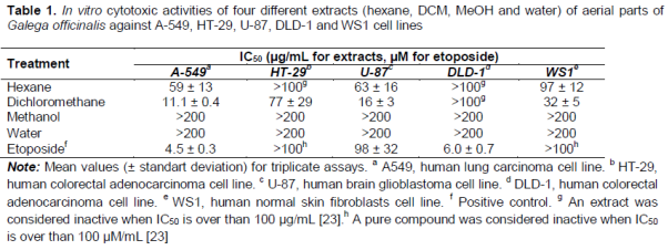
Results: DCM extract showed strong cytotoxic activity against lung adenocarcinoma and brain glioblastoma cell lines, with IC50 (concentration inhibiting 50 % of cell growth) values of 11 ± 0.4 and 16 ± 3 µg/mL, respectively. The hexane extract showed moderate anticancer activity against the same cell lines (59 ± 13 and 63 ± 16 µg/mL, respectively). DCM extract also showed significant anti-inflammatory activity, inhibiting NO release by 86.7 % at 40 µg/mL in lipopolysaccharide (LPS) - stimulated murine RAW 264.7 macrophages. Of all test extracts, the methanol extract of G. officinalis showed the highest antioxidant activity with 2.33 ± 0.09 µmol Trolox/mg , 7.10 ± 0.9 g tannic acid equivalent (TAE), and IC50 of 44 ± 4 µg/mL.
Conclusion: The findings of this study suggest that DCM extract may possess anticancer effect against lung adenocarcinoma and brain glioblastoma, as well as serve as an anti-inflammatory agent.
Introduction
Galega officinalis L., which belongs to Leguminosae family, is generally known as goat’s rue and is a native plant in Southeastern Europe, Middle East, and Western Asia. It grows wild in damp and low-lying areas [1,2]. Aerial parts of this crop is widely used as a functional remedy against malignant fever, inflammation, parasitic infection, worms, and diabetes in Europe [3,4]. It has also been used to increase breast milk production in folk medicine [5,6]. This plant was described as a diuretic [3] and anti-diabetic crop, having the capacity to reduce blood sugar levels [3,7]. Goat rue contains some secondary metabolites such as alkaloids (including galegine), saponins, flavonoids (flavonol triglycosides, kaempferol, and quercetin) [14], tannins [3] and fatty acids (α-linolenic acid, palmitic acid and linoleic acid) [15] and phytoestrogens (medicarpin, and sativan) [15,16]. Galegine, a compound isolated from leaves of G. officinalis [3] and metformin, a synthetic form of galegine reduce blood sugar levels and find in many anti-diabetic drugs as an active molecule [7-9].
Atanasov and Spasov [10] have shown that the aqueous extract and the gel-filtered fractions from the G. officinalis have inhibitory activity on platelet aggregation. The antibacterial activities of G. officinalis have been shown against to Gram positive (Staphylococcus aureus and Staphylococcus epidermidis) and gram-negative (Serratia marcescens, Salmonella typhimurium, Pseudomonas aeruginosa, Proteus vulgaris and Eschericia coli) bacteria [2,12]. There are also some studies demonstrating toxicity of aerial parts of G. officinalis in sheep [12,13] and in rats [6].
The present study was carried out to evaluate the cytotoxic, anti-inflammatory, and antioxidant activities of four different fractions obtained from the aerial parts of Galega officinalis.
Methods
Plant material and extraction procedure
The aerial parts of the goat rue (Galega officinalis L.) were obtained from Abant Izzet Baysal University Campus, Bolu, Turkey in July. Recognition of the plant species was carried out by Prof. Dr. Arzu Ucar Turker using “Flora of Turkey and the East Aegean Islands” [1], and a voucher specimen (collection no. AUT-1912) was kept at the Abant Izzet Baysal University (AIBU) Herbarium, Bolu, Turkey.
The collected plant materials were cleaned and oven dried at 40 oC for seven days. The dried aerial parts of G. officinalis were powdered and 100 g dried weight of plant material was successively extracted with 800 mL hexane (at 65 - 70 oC), 600 mL dichloromethane (at 55 - 60 oC), 600 mL methanol (at 60 oC) and 300 mL water (at 80 oC) by using soxhlet apparatus for 18 hrs. The four different fractions of G. officinalis were filtered with Whatmann No 1 filter paper. The fraction solvents were concentrated by a rotary evaporator under low vacuum at 40 oC to dryness. The yield of the extracts (w/w) were 1.9 % for hexane, 0.38 % for DCM, 10.2 % for MeOH and 10.36 % for water fractions. The dried extracts were stored in the dark at -20 oC. For biological activity assays, the crude extracts were dissolved in sterile dimethyl sulfoxide (DMSO) in order to obtain a final concentration of 80 mg/mL.
Cell lines and culture conditions
Human lung carcinoma A-549, human colorectal adenocarcinoma HT-29, human brain glioblastoma U-87, colon adenocarcinoma DLD-1, normal skin fibroblast WS1 and murine macrophage RAW 264.7 cell lines were provided from the American Type Culture Collection (ATCC, Manassas, VA, USA). The used cell lines were augmented in Dulbecco’s minimum essential medium (DMEM) with Earle’s salts (Mediatech Cellgro, Herndon, USA),which was completed with 10 % fetal bovine serum (FBS; Hyclone, Logan, USA), a solution of vitamins, sodium pyruvate and non-essential amino acids (all at 1:100 v/v dilution of supplied solutions), streptomycin (100 µg/ mL), and penicillin (100 IU/mL) (Mediatech Cellgro, VA). A humidified atmosphere at 37 °C in 5 % CO2 was available for incubation of all cells.
Screening for cytotoxic activity
Growing cells were exponentially located in 96-well microplates (Costar, Corning Inc.) at a density of 5 × 10³ cells per well in 100 µL of culture medium and were led to adhere for 16 h before application. All G. officinalis extracts were made in a series of eight two fold dilutions (0 – 200 µg/mL). Then, 100 µL of raising levels of tested extracts were put into to three wells. The highest level of DMSO in the culture medium was sustained at 0.5 % (v/v) to eliminate solvent toxicity. The cell cultures were cultured for 48 h in the presence or absence of G. officinalis extracts. Etoposide (≥ 98 %, Sigma–Aldrich) was utilized as a positive control for the relevant comparison. The resazurin reduction test was used to detect the cell viability [17]. Fluorescence was read for the metabolic activity of cells in each well by Fluoroskan Ascent FL Thermo plate reader (Labsystem). Survival percentages of cells were detected as the fluorescence in experimental wells compared to that in control wells after subtraction of blank values. After reading the resazurin test, cells were treated for cellular DNA assay with Hoechst dye 33342. After washing with PBS solution, dried cells were kept at -80 °C until the Hoechst assay was carried out [18]. Cytotoxicity data were represented by using means ± standard deviation and expressed by the concentration that inhibited 50 % of cell growth (IC50). Each concentration of four different extracts from G. officinalis was measured three times.
Determination of anti-inflammatory activity
Growing cells were exponentially plated at a density of 75 × 103 cells per well in 96-well microplates (BD Falcon) in 100 µL of cell culture medium and were allowed to cohere overnight. Each G. officinalis extract was made in a series of 8 two fold dilutions (final concentrations ranging from 0 to 160 µg/mL) in DMSO and medium culture. The cells were either applied or not with positive control N(G)-nitro-L-arginine methyl ester hydrochloride (L-NAME, ≥ 98 %, Sigma–Aldrich), or raising levels of G. officinalis extracts on 3 wells.
Tested cells were then induced with 100 µg/mL of LPS and cultured at 37 °C, 5 % CO2 for 24 h. After 24 h, cell-free supernatants were gathered and NO level was instantly detected by means of the Griess reaction with a small amount of modifications [19]. Furthermore, 100 µL aliquots of cell supernatants were cultured with an equal volume of Griss reagent (50 µL of 1 % sulfanilamide in 2.5 % H3PO4 and 50 µL of 0.1 % N-1-naphtyl-ethylenediamine dihydrochloride in water) at 22 ± 2 °C for 20 min. Absorbance at 550 nm was then determined via an automated 96-well Varioskan Ascent plate reader (Thermo Electron) and the presence of nitrite was measured by comparison with sodium nitrite (NaNO2) standard curve. Macrophage survival was measured by means of the resazurin reduction test.
Assessment of oxygen radical absorbing capacity (ORACFL) assay
The antioxidant properties of G. officinalis extracts were determined by using oxygen radical absorbance capacity (ORAC) assay that detects their scavenging capacity against peroxyl radicals. The experimental method was modified from the process defined by Ou et al [20]. Shortly, the ORAC assay was made using 96-wells microplates and a Fluoroskan Ascent FlTM microplate reader (Labsystems, Milford, MA, USA). A positive control standard was trolox, a water-soluble analog of vitamin E.
The study was managed at 37.5 °C and pH 7.4 with a blank sample. The final data were determined by calculating the net areas under the fluorescein decay curves between the blank and the treatments. ORAC values were represented in µmol of Trolox equivalents/mg dry weight of sample (µmol TE/mg dw).
Total phenolic assay
The total phenolic content was analyzed by using the Folin-Ciocalteu reagent according to the procedure reported by Singleton and Rossi [21]. The experiment was performed 3 times, and the data were represented in g TAE/100 g dried mass.
Antioxidant cell assay
The antioxidant activity was also performed using the 2′,7′-dichlorofluorescin-diacetate (DCFH-DA) assay as defined by Girard-Lalancette et al [22], with some minor changes. Growing WS1 cells were exponentially plated in 96-wells microplates at 10 × 103 cells per well and maintened for one day at 37.5 °C and 5 % CO2. The WS1 cells were rinsed with 150 µL Hank’s balanced salt solution (HBSS) at pH 7.4 and cultured for 30 min with 100 µL HBSS (pH 7.4) including 5 µM DCFH-DA (Sigma–Aldrich). The WS1 cells were rinsed again with 150 µL HBSS. To evaluate the cellular antioxidant activity, the cells were cultured either with an increasing level of four different extracts from G. officinalis, quercetin or trolox, in the absence or presence of 200 µM tert -butylhydroperoxide (t- BuOOH). After t- BuOOH treatment, fluorescence was determined instantly. Again 90 min later, it was read by using an automated 96-well plate reader (Fluoroskan Ascent Fl, Thermo-Labsystems) at an excitation wavelength of 485 nm and an emission wavelength of 530 nm.
Statistical analysis
Data were presented as mean ± SD and analyzed using SPSS software (version 22.0, SPSS Inc, Chicago, IL, USA) for Windows by one-way ANOVA using Duncan’s Multiple Range Tests. Differences were considered statistically significant at p ≤ 0.05.
Results
The results for antiproliferative activities of four different extracts from G. officinalis against A-549, HT-29, U-87, DLD-1, and WS1 cell lines were represented in . In this experiment, DCM and hexane extracts were found to display from strong to moderate cytotoxicities when IC50 values were between 11 and 100 µg/mL. DCM extract of G. officinalis showed the highest cytotoxic activity against to A-549 (11 ± 0.4 µg/mL) and U-87 (16 ± 3 µg/mL) cancer cell lines (). The hexane extract showed a moderate anti-cancer activity against to A-549 and U-87 cell lines, with IC50 values of 59 ± 1 and 63 ± 16 µg/mL, respectively (). However, the MeOH and water extracts were inactive against to A-549, HT-29, U-87, and DLD-1 with an IC50 value higher than 100 µg/mL ().
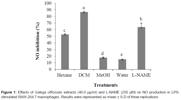
The anti-inflammatory activity of the extracts was observed by assessing their ability to prevent cellular NO production, which is an endogenous free radical species. As shown in , the DCM extract showed stronger inhibitory effect on LPS-induced NO secretion with 86.7 % inhibition than hexane (52.9 % inhibition), methanol (17.8 % inhibition) and water (16.2 % inhibition) extracts of G. officinalis observed at 40 µg/mL concentration. Relatively, the L-NAME prevented NO release by 63.9 % at 250.0 μM (67.4 μg/mL). The anti-inflammatory properties were evaluated at non-cytotoxic concentrations. To investigate the effects of G. officinalis extracts on the viability of murine macrophages (RAW 264), we carried out resazurin assay. When we treated with the extracts at concentrations of 1.25, 2.5, 5, 10, 20, 40, 80, 100 µg/mL, it had no effect on cell viability.
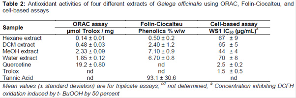
The incubation of murine macrophages with G. officinalis extracts and 10 μg/mL LPS also did not show any cytotoxicity (data not shown). In , the current findings demonstrated that the MeOH and water extracts were moderate antioxidant, with ORAC values of 2.33 ± 0.09 and 1.85 ± 0.12 µmol TE/mg of dry weight, respectively. The hexane and DCM extracts were low antioxidant, with ORAC values, respectively, of 0.14 ± 0.00 and 0.48 ± 0.03 µmol TE/mg of dry mass. The ORAC value of standard phenolic compound used as a positive control (quercetine) was 19.20 ± 0.80 µmol TE/mg ().
The data for the total phenolic content () showed that the MeOH and water extracts of G. officinalis had low levels of phenolic contents (7.10 ± 0.9 and 6.70 ± 0.8 g TAE/100 g of extract, respectively). Similarly, the hexane (0.50 ± 0.2 TAE/100 g) and DCM (2.40 ± 1.2 g TAE/100 g) extracts contained low concentration of phenolic compounds.
The cell-based assay results (expressed as IC50) of t-BuOOH-induced oxidation of 2′,7′-dichlorofluorescin (DCFH) indicate that quercetin and trolox, used as positive controls, had IC50 of 2.5 ± 0.2 and 1.5 ± 0.5 μg/mL, respectively (). Hexane (67 ± 9 µg/mL), DCM (65 ± 5 µg/mL), MeOH (44 ± 4 µg/mL), and water (70 ± 8 µg/mL) extracts prevented tBH-induced oxidation of DCFH by 50 %.
Discussion
The DCM extract of G. officinalis has promising anticancer activity against A549, HT-29, and U-87 with IC50 < 100 µg/mL. An extract is accepted as active against cancer cell lines when IC50 value is < 100 µg/mL [23,24]. The anticancer activity of DCM and hexane extracts can be attributed to the alkaloids (galegine) or flavonoids (kaempferol, quercetin, medicarpin and sativan) found as dominant substances in goat rue [14]. According to one study [14], sativan and medicarpin as phytoestrogens isolated from the leaves of goat rue were found to be cytotoxic against human breast cancer cell lines. Morever, the anti-mutagenic, anti-inflammatory, anti-viral, and anti-carcinogenic activities of phytoestrogens have also been demonstrated formerly [14].
Although the aerial parts of G. officinalis have been widely used for the treatment of inflammatory diseases in traditional medicine [3-4], the anti-inflammatory activity of this medicinal plant has not been scientifically verified up to now. This is the first report about the anti-inflammatory activity of G. officinalis. Nitrit oxide (NO) is an important regulator of body homeostasis in animals and high amounts of NO causes many serious inflammatory diseases [25]. For this reason, prevention of NO generation may be an effective approach for the remedy of various inflammatory diseases [25]. The anti-inflammatory activity of the extracts was considered using LPS-induced RAW 264.7 macrophages. L –NAME was used as a positive control, because it inhibits the formation of NO in LPS-stimulated murine RAW 264.7 macrophages [26]. A prerequisite for anti-inflammatory studies is that the extract or substance being tested should not have cytotoxic activity toward murine macrophage RAW 264.7 cells. Inhibition of NO secretion in murine macrophage RAW 264.7 cells is caused by two effects: by anti-inflammatory property or decreasing the cell viability of the murine macrophages [19]. indicated that the inhibition rates of NO production by the DCM extract of G. officinalis was 86.7 % at the concentration of 40 μg/mL. The cell viability data represents the strong inhibitory effect of the DCM extract on NO secretion is not due to its cytotoxicity (data not shown). The strong anti-inflammatory activity of the DCM extract of G. officinalis may be attributed to the alkaloid or flavonoid constituents of this plant [14].
The antioxidant activities (in vitro and ex vivo) of different G. officinalis extracts have not been previously investigated in the literature. Among the tested extracts, MeOH extract had the best phenolic content, ORAC value, and IC50 value in cell-based assay. The revealed antioxidant activity can be explained by the presence of some phenolic substances in the extract ingredient. Although DCM extract had the best anticancer and anti-inflammatory activities, it includes low amounts of trolox and total phenolic content. At the same time, hexane extract showed potent anticancer activity against A549 and U-87 (IC50 of 59 ± 13 µg/mL and 63 ± 16 µg/mL), and anti-inflammatory activity on LPS-induced NO secretion with 52.9 % inhibition at 40 µg/mL concentration. This is possibly due to the presence of some specific phenolic compounds, terpenoids or alkaloids in the DCM and hexane extracts. The various amount of secondary metabolites present in G. officinalis extracts could be explained by the different polarity of the extraction solvents (hexane, DCM, methanol, and water, from non-polar to polar, respectively).
Conclusion
The anticancer, anti-inflammatory, and antioxidant activities for four different solvent extracts from Galega officinalis revealed that the DCM and hexane extracts from the aerial parts were active against human lung and brain glioblastoma carcinoma cell lines. DCM extract also exhibited remarkable anti-inflammatory activity. On the basis of the findings of this study, there seems to be some justification for the use of the aerial parts of G. officinalis in traditional medicine as an anti-inflammatory agent. Future studies are required to isolate the chemical constituents and to estimate the inhibitory effect of individual compounds of G. officinalis on cancer and inflammation diseases.
Declarations
Acknowledgement
References
Archives

News Updates